The human skull is made of 22 separate bones fused into one protective structure — a count established in classic anatomical texts like Gray’s Anatomy and confirmed in modern medical references. This guide breaks down every bone, the named joints between them, the muscles attached to the skull, and what’s actually happening beneath the bone to protect the brain — with labeled diagrams throughout — everything you need to understand the anatomical skull in one place.
What Is the Anatomical Skull?
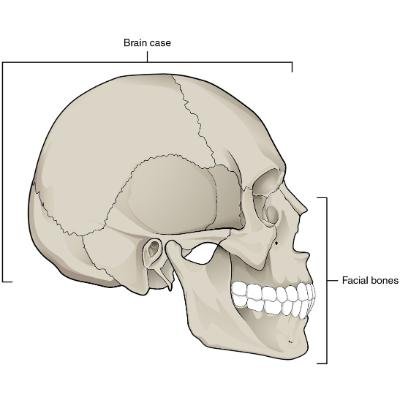
The skull serves two jobs at once: protecting the brain and giving structure to the face. Anatomists divide it into two connected sections: the neurocranium (the bony case around the brain) and the viscerocranium, or facial skeleton — a division that, per the Wikipedia entry on the human skull, reflects each section’s distinct embryological origin rather than just its final shape.
The Cranium and Facial Skeleton
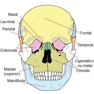
The cranium is made of 8 bones: the frontal, two parietal, two temporal, occipital, sphenoid, and ethmoid bones. In infancy, these bones aren’t fused yet. Instead, they’re separated by soft, unossified gaps called fontanelles, which close on their own timeline over the first two years of life. As a child grows, the fontanelles narrow into rigid but distinct joints called sutures.
The facial skeleton is the remaining 14 bones: the two nasal bones, two maxillae (upper jaw), two zygomatic bones (cheekbones), two palatine bones, two lacrimal bones, two inferior nasal conchae, the vomer, and the mandible (lower jaw). According to the anatomy reference maintained on the NCBI Bookshelf, the mandible is the only bone in the skull that forms a true, independently movable joint. Every other bone, in contrast, locks in place once its sutures fully ossify.
Skull anatomy also has applications outside clinical medicine. In biological anthropology and forensic science, suture patterns, cranial capacity, and bone proportions are used to study population history and assist in individual identification — the same underlying anatomy covered in our guide to elongated skulls, where entire cultures intentionally reshaped this structure in infancy.
If you’d rather explore the structure interactively than read diagrams, our 3D skull model lets you rotate and zoom through each bone.
Cranial Bones of the Anatomical Skull (Labeled)
The 8 cranial bones of the anatomical skull work together to form a protective vault around the brain:
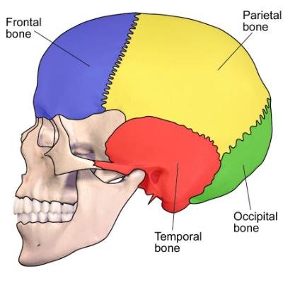
- Frontal bone — forms the forehead and the upper part of the eye sockets.
- Parietal bones (2) — form the sides and roof of the cranium, meeting at the top of the skull.
- Temporal bones (2) — sit above the ears and house the structures involved in hearing. Each one subdivides into four parts: petrous, squamous, zygomatic, and mastoid. The petrous portion, specifically, houses the inner ear.
- Occipital bone — forms the back and base of the skull; contains the foramen magnum, the opening where the spinal cord connects to the brain.
- Sphenoid bone — a butterfly-shaped bone at the base of the skull that articulates with nearly every other cranial bone, making it a key point of structural stability.
- Ethmoid bone — a lightweight bone between the eyes that forms part of the nasal cavity and eye sockets.
Facial Bones (Labeled)
The 14 facial bones give the face its shape and handle the mechanical work of eating and speaking:
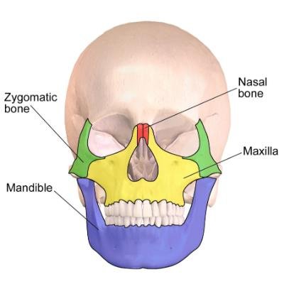
- Nasal bones (2) — form the bridge of the nose.
- Maxillae (2) — hold the upper teeth and form part of the eye sockets.
- Zygomatic bones (2) — the cheekbones; also help shield the eyes.
- Palatine bones (2) — form the back of the hard palate, separating the nasal and oral cavities.
- Lacrimal bones (2) — the smallest bones in the skull; sit in the inner corner of the eye socket and help drain tears.
- Inferior nasal conchae (2) — thin, curled bones inside the nasal cavity that add surface area for warming and filtering air.
- Vomer — a single bone forming part of the wall dividing the two nasal passages.
- Mandible — the lower jaw, and the only bone in the skull that moves independently.
Cranial Sutures and Landmarks
The joints between cranial bones aren’t simple seams — they’re named, clinically significant structures. Where they intersect, they form landmarks still used throughout medicine today.
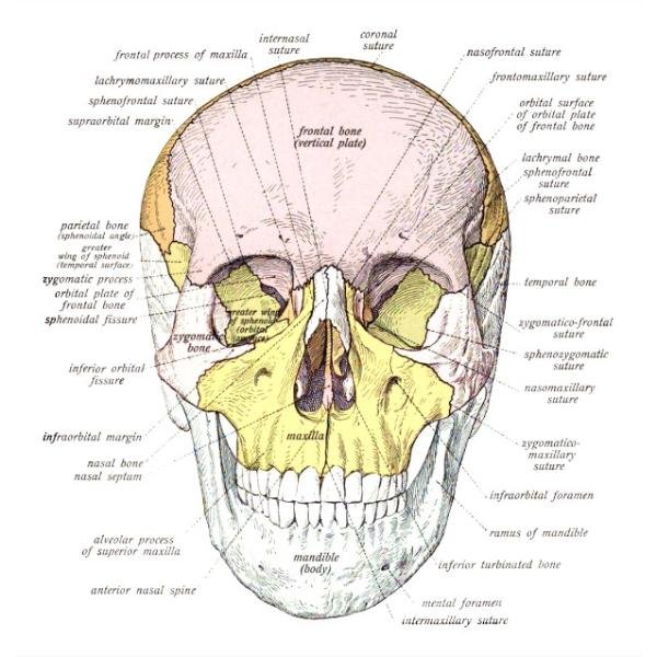
The four major sutures:
- Coronal suture — runs ear-to-ear, separating the frontal bone from the two parietal bones. The name comes from the Latin corona (“crown”), referring to the position where a crown would sit.
- Sagittal suture — runs front-to-back along the midline, joining the two parietal bones to each other.
- Lambdoid suture — an inverted-V-shaped suture at the back of the skull, joining the parietal bones to the occipital bone, named for its resemblance to the Greek letter lambda (Λ).
- Squamous suture — joins the temporal bone to the parietal bone above the ear on each side.
Where sutures cross, they form named reference points: the bregma (coronal and sagittal sutures meet) and the lambda (sagittal and lambdoid sutures meet). On the side of the skull, the pterion marks where the frontal, parietal, temporal, and sphenoid bones all converge — clinically important because the bone is thin there and the middle meningeal artery runs directly beneath it.
Suture Closure and Bone Formation
Sutures don’t all close at the same age. The anatomy reference on the NCBI Bookshelf documents that the sagittal suture typically ossifies first, around age 22, followed by the coronal suture, then the lambdoid suture around age 26 — while the squamous suture can remain unfused until as late as age 60.
Bone formation across the skull isn’t uniform either. The flat bones of the cranial vault and face develop through intramembranous ossification, meaning bone forms directly, without a cartilage template. The bones at the skull base, however — the sphenoid and ethmoid — develop through endochondral ossification instead, where bone replaces an earlier cartilage structure. This second process is, in fact, the same one that forms most of the rest of the skeleton.
Cranial Fossae and Brain Protection
The floor of the cranial cavity steps down in three distinct basins — the anterior, middle, and posterior cranial fossae — that cradle different parts of the brain at different heights, a structure described in detail in classic osteology texts derived from Gray’s original 1918 Anatomy of the Human Body.
- The anterior cranial fossa, formed by the frontal, ethmoid, and sphenoid bones, sits highest and supports the frontal lobes.
- The middle cranial fossa, formed by the sphenoid and temporal bones, sits lower and houses the temporal lobes, along with the pituitary gland at its midline.
- The posterior cranial fossa, the largest and deepest, is formed mainly by the occipital and temporal bones and holds the cerebellum and brainstem. It contains the foramen magnum, where the spinal cord exits the skull.
Directly against the bone, three thin membranes called the meninges wrap the brain: the dura mater, arachnoid mater, and pia mater. Between the arachnoid and pia mater sits the subarachnoid space. This space is filled with cerebrospinal fluid, which cushions the brain against impact.
Muscles Attached to the Anatomical Skull
The anatomical skull anchors muscles that handle facial expression, chewing, and head movement.
Facial expression muscles attach directly to skin and bone, rather than to other muscles. The frontalis raises the eyebrows, for example, while the zygomaticus major pulls the corners of the mouth up when smiling. This direct attachment is part of why facial expressions can be so precise and varied.
Chewing muscles do the mechanical work. The masseter runs from the cheekbone to the mandible and, relative to its size, is one of the strongest muscles in the human body. The temporalis, a fan-shaped muscle on the side of the skull, assists the masseter and helps stabilize the jaw.
Head movement relies on the sternocleidomastoid, which connects the skull to the collarbone and breastbone and lets the head turn, tilt, and nod.
Curious about the cultural and historical side of skulls too? Visit our skull information hub for anatomy guides, history, myths, and our full skull-themed shop.
Anatomical Skull FAQ
The anatomical skull has 22 bones in total: 8 cranial bones that form the protective case around the brain, and 14 facial bones that shape the face. This count, standard since Gray’s Anatomy, excludes the ear ossicles and the hyoid bone, which sit in the head and neck region but aren’t classified as part of the skull itself.
The mandible (lower jaw). Every other bone in the skull is fixed in place by sutures once fully ossified.
Three main groups: facial expression muscles (like the frontalis and zygomaticus major), chewing muscles (the masseter and temporalis), and the sternocleidomastoid, which connects the skull to the neck and enables head movement.
The large opening in the occipital bone, at the base of the posterior cranial fossa, where the spinal cord passes through to connect with the brain.
Sutures are the fibrous joints between skull bones. They stay flexible through childhood to allow brain growth, then progressively ossify. Per the NCBI Bookshelf’s anatomy reference, the sagittal suture typically closes first, around age 22. The coronal suture follows, then the lambdoid suture around age 26. The squamous suture, however, can remain unfused until as late as age 60.
A newborn’s cranial bones aren’t fused at all. Instead, they’re separated by open fontanelles, which allow the head to pass through birth and the brain to grow rapidly in the first two years. Our newborn skull guide covers that anatomy specifically.
Final Thoughts
The anatomical skull’s twenty-two bones, four major sutures, three cranial fossae, and a handful of muscles work together to do two jobs at once. They protect one of the body’s most vital organs, and they give the face its structure and movement. As a result, it’s an arrangement precise enough that clinicians still navigate it by named 19th-century landmarks like the bregma and pterion. It’s also consistent enough that the 22-bone count established in 19th-century anatomical texts still holds up against modern medical references today.
References / Further Reading
- Wikipedia — Skull
- NCBI Bookshelf, StatPearls — Anatomy, Head and Neck, Skull
- NCBI Bookshelf, StatPearls — Anatomy, Head and Neck, Coronal Suture
- Cleveland Clinic — Skull (Cranial) Sutures: What They Are & Anatomy
- Kenhub — Skull joints and sutures: Anatomy and functions